Our imaging capabilities allow us to look inside your pet without invasive procedures. From broken bones to soft tissue masses, digital X-rays and ultrasound give us critical information quickly — helping us reach an accurate diagnosis and start treatment sooner.
Digital Radiography
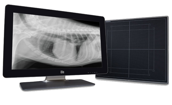
Our digital X-ray system produces high-resolution images instantly — no waiting for film to develop. Digital radiography uses less radiation than traditional X-rays and allows us to zoom in, adjust contrast, and share images with specialists when needed.
Bone & Joint Evaluation
Fractures, arthritis, hip dysplasia, and skeletal abnormalities.
Chest X-Rays
Heart size, lung disease, pneumonia, and fluid in the chest.
Abdominal X-Rays
Foreign body ingestion, bladder stones, intestinal obstruction, and organ size.
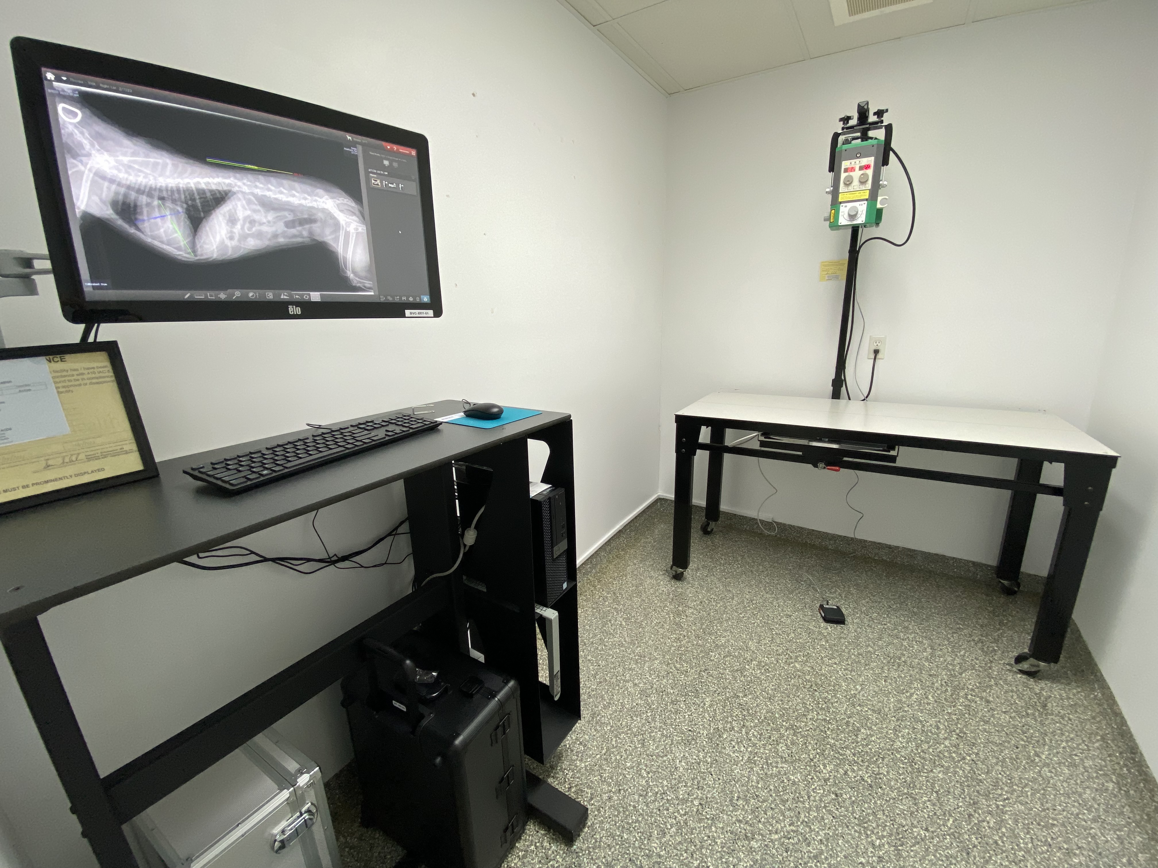
Our X-Ray Suite
Our dedicated X-ray room is equipped with a mobile X-ray unit on a movable arm and a large digital display so we can review images with you in real time.
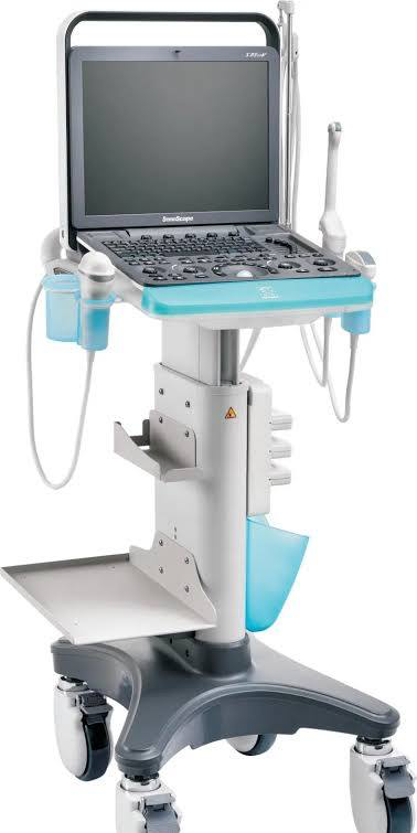
Ultrasound
Ultrasound uses sound waves to create real-time images of your pet’s internal organs and soft tissues. It’s particularly valuable for evaluating the heart, liver, kidneys, spleen, and bladder — and for guided biopsies or fluid sampling.
Cardiac Evaluation
Assess heart wall motion, valve function, and fluid around the heart.
Abdominal Imaging
Evaluate organ size, texture, and detect masses, cysts, or fluid.
Guided Sampling
Ultrasound-guided biopsies and fluid aspiration for accurate diagnosis.